In facial nerve palsy, the corner of the mouth of the paralyzed side hangs down like a suspension bridge where the steel cable is not fixed on one side. To correct the hanging, the corner of the mouth can be "hung" on the zygomatic arch (the prominent bone below the eye symbolically serves as a "support"). For this purpose, the body's own connective tissue from the thigh or tendon tissue from the forearm can be used. The body's own tissue is particularly suitable because, unlike artificial materials, it does not cause rejection reactions and is more similar to the original tissue in terms of elasticity and tension[1, 2]. The most commonly used for this purpose is the fascia lata (flat and at the same time tight connective tissue from the thigh). This is obtained from the lateral thigh, for which only small skin incisions are required[3]. Functionally, the tissue at this point can be dispensed with. This means that there are no functional deficits of the limb, such as when walking. After appropriate processing and preparation, the patient's own tissue is inserted in smaller strips via an inconspicuous face-lift access. It is fixed to the corner of the mouth and the mouth ring muscle and is suspended from the zygomatic arch. In this way, flaccid tissue in the cheek,
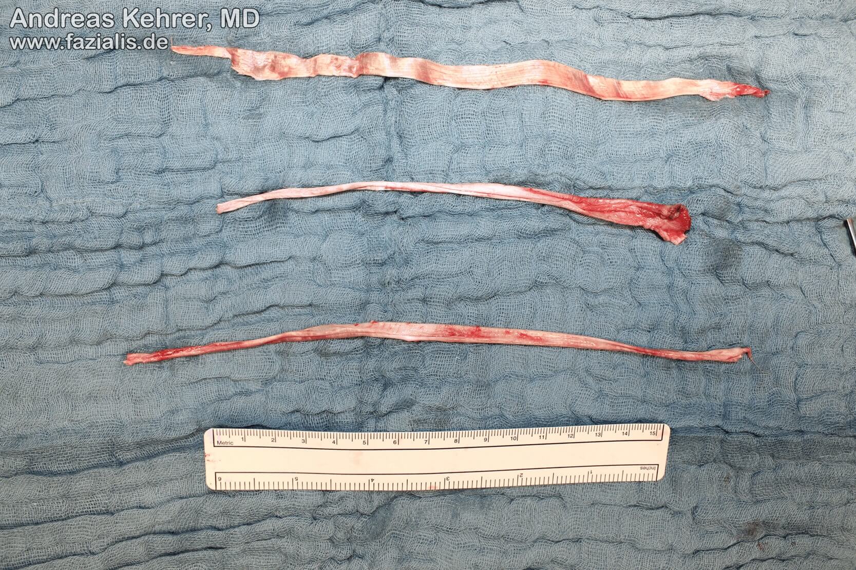
lip and mouth corner areas can be tightened significantly. The tension is individually adapted to the patient in order to achieve the best possible symmetry of rest. Patients also benefit from improved oral continence and pronunciation[4].
This procedure is suitable, for example, for patients who, for certain reasons, are not eligible for dynamic reconstruction (resuscitation of facial expression by muscle grafting or motor replacement plastic surgery).
However, the operation can also be combined with dynamic reconstruction, e.g. free muscle transplantation[4]. This often achieves the best aesthetic and functional results in middle-aged patients. While the fascia lata's own tissue creates symmetry at rest, i.e. when the patient relaxes his or her face, the free functional muscle ensures symmetry with the opposite side when smiling or laughing, for example.
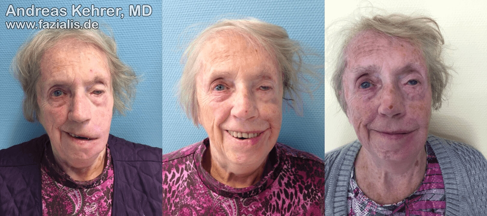
87-year-old female patient: the stigma of facial paralysis due to asymmetry becomes particularly clear when smiling and often leads to tendencies to withdraw from society, as with this otherwise very cheerful and sociable patient. The outer clothing, which is constantly soiled by the uncontrollable flow of saliva over the hanging corner of the mouth on the left, often plays a role (left picture). After facial reconstruction, the patient succeeds in having a symmetrical smile with open lips and "showing teeth". By restoring oral continence, the blouse is free of saliva deposits. The eyelid correction of the left eye was not completed at this time (center). After raising the eyebrows, insertion of an adapted platinum weight into the upper eyelid and correcting the lower eyelid, vision in the left eye is possible again. The pupil is no longer obscured by the upper eyelid. The patient has clearly regained self-confidence and increased her postoperative social activities enormously.
Sources:
[1] Morgan AS, McIff T, Park DL, Tsue TT, Kriet JD. Biomechanical properties of materials used in static facial suspension. Arch Facial Plast Surg. 2004;6(5):308-10.
[2] Constantinides M, Galli SK, Miller PJ. Complications of static facial suspensions with expanded polytetrafluoroethylene (ePTFE). Laryngoscope. 2001;111(12):2114-21.
[3] Leckenby JI, Harrison DH, Grobbelaar AO. Static support in the facial palsy patient: a case series of 51 patients using tensor fascia lata slings as the sole treatment for correcting the position of the mouth. J Plast Reconstr Aesthet Surg. 2014;67(3):350-7.
[4] Rose EH. Autogenous Fascia Lata Grafts: Clinical Applications in Reanimation of the Totally or Partially Paralyzed Face. Plastic and Reconstructive Surgery. 2005;116(1):20-32.