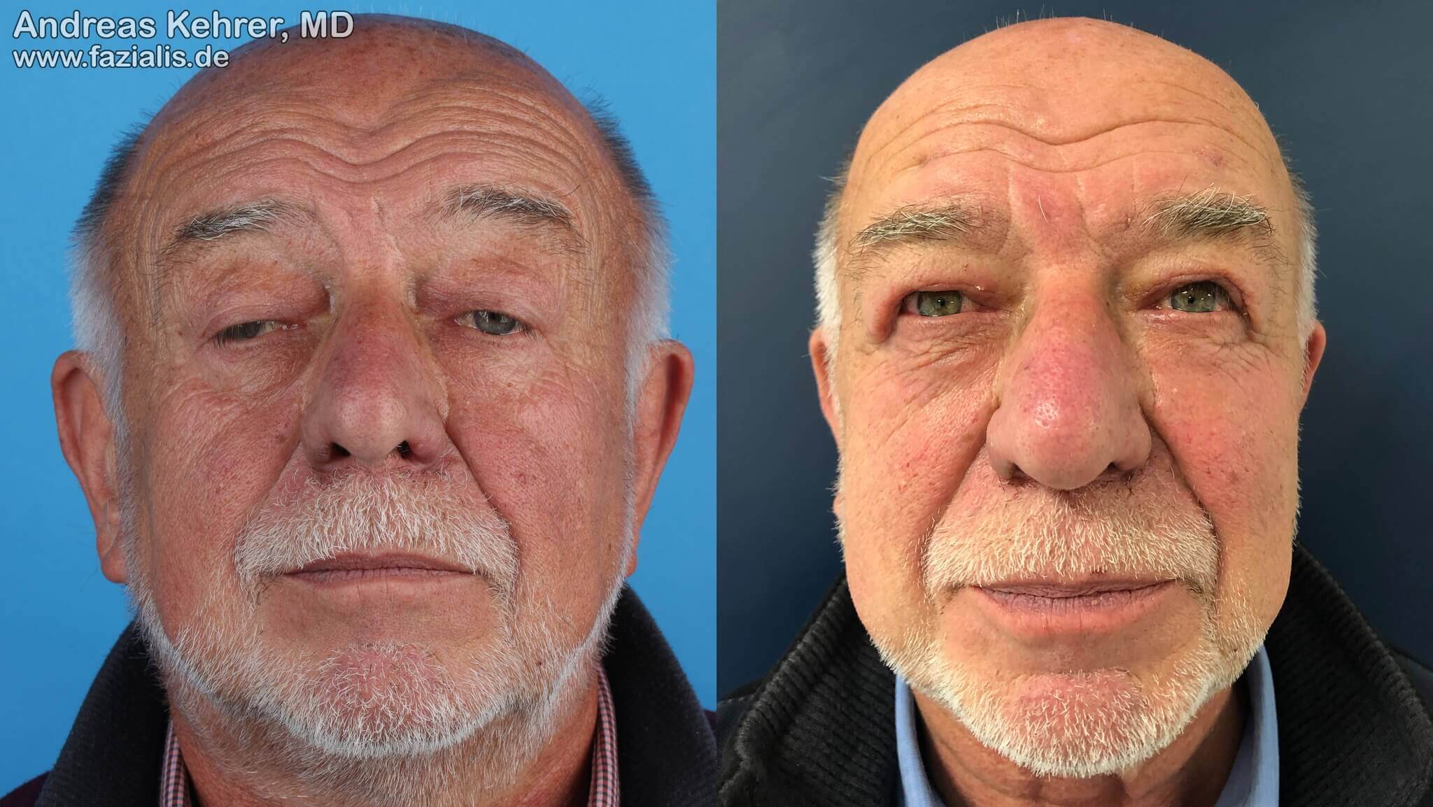
Congenital eyelid ptosis in a 70-year-old man. The levator muscle responsible for the eyelid lift is paralyzed on both sides and the patient can hardly open his eyes. The palpebral aperture is severely reduced on both sides, the patient's field of vision, especially in upward gaze, is clearly restricted. The patient tries to compensate the visual field deficit as much as possible by constant tension of the forehead muscles, which has already led to deep furrows in the forehead area. In order to be able to look horizontally ("straight view"), the patient has to constantly place his head in the neck in order to get along reasonably in everyday life (left picture). He already complained of neck pain. The gentleman was reconstructed on both sides with a (divided) fine tendon transplanted from the left forearm - a frontalis sling operation (right picture). The tendon strips running under the skin now connect the upper eyelids directly with the forehead muscle and, with their static component as a suspension sling, ensure a much more favorable position of the upper eyelids at rest. The palpebral aperture has been enormously improved and a horizontal view is now effortless. If the gentleman now also raises his eyebrows spontaneously, he can open his eyes even further via the new connection through the tendon strips (dynamic component). In the right picture, four weeks after the operation, there is still moderate swelling of the upper eyelids on both sides. A closer look reveals small, minimally red scars in the forehead area above the eyebrows as well as a residual thread on the right upper lid. In a few weeks, none of the operation scars will be visible and the swelling will be completely gone. If you look closely, you will see that the wrinkles in the forehead area have already improved in the right picture - the patient apparently no longer tenses the forehead muscle constantly, the furrows have become more flat.