Anatomy (ancient Greek: ἀνά aná "auf" and τομή tomé "Schnitt") is the science of the "construction" of an organism. In medicine, this study is divided into macroscopic anatomy (coarse structure of organs and tissues) and microscopic anatomy (structures which are evaluated with the help of microscopes).
Nerves are parallel nerve fibers that are surrounded by connective tissue and can conduct electrochemical impulses to peripheral organs, muscles and the central nervous system. A nerve cell basically consists of the soma (cell body, cell nucleus with dendrites), the axon and the synapses (axon terminals). A single nerve fiber is surrounded by the "endoneurium". Several nerve fibres are surrounded by the "perineurium" and together form a nerve fascicle. Nerve fascicles are in turn grouped by the epineurium to form the anatomical structure of a peripheral nerve.
In order to pass information from one cell to the next, the so-called "synaptic cleft" must be bridged. This represents a 20 to 30 nm wide gap between two cells.[1] The synaptic cleft forms the synapse together with the axon end button of the transmitter cell and the dendrite of the receiver cell.
The axon end button contains small vesicles, which in the case of chemical synapses contain messenger substances such as the neurotransmitters adrenaline and dopamine. When an electrical impulse reaches the end button, the vesicles fuse with the presynaptic cell membrane, i.e. the one in front of the synaptic cleft, and the messenger substances are released into the synaptic cleft. The electrical impulse is converted into a chemical signal.
At the cell membrane of the recipient cell, i.e. the postsynaptic membrane, there are docking sites (receptor molecules) for the messenger substances. When a transmitter binds to a receptor molecule, an electrical signal is again triggered in the recipient cell, which can propagate along the cell. In this way, nerve impulses are transmitted from cell to cell. Exchange rates of 1 to 100 m/s are achieved.[2] The speed of nerve conduction depends, among other things, on whether the nerve is surrounded by a fat-rich biomembrane (myelin sheath).
We distinguish between multipolar (for sensory and motor tasks), unipolar (only sensory perception), bipolar (reception and transmission of signals from rods and cones in the cornea of the eye) and pseudounipolar (with only one extension from the soma) nerve cells. Furthermore, afferent (conducting signals from sensory cells to the central nervous system) and efferent (conducting signals from the central nervous system to muscles and glands) neurons are differentiated. Furthermore, central (CNS) and peripheral parts (PNS) of the nervous system can be distinguished. The cranial nervous system is a special form of PNS. The PNS can be divided into somatic (arbitrary) and autonomous (vegetative) parts.
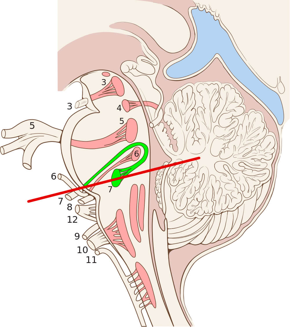
Figure 1
Schematic representation of the cranial nerve nuclei of all 12 cranial nerves in the brain stem. Usually the cranial nerves are assigned Roman numerals, so that for example the facial nerve is called the VII. cranial nerve (here 7, in green) and the N. massetericus is called the Vth cranial nerve (here 5). In this schematic drawing, the topographical proximity of both cranial nerve nuclei (VII and V) becomes clear, which is held responsible for a certain ability of both cranial nervous systems to communicate with each other. This is potentially more important when nerve cells (neurons) of the V. Brain nerves after a nerve transfer (microsurgical nerve rearrangement in the face) are to take over original tasks of nerve cells of the VII brain nerves, i.e. for example, to trigger the muscles to smile. If this process, which is accelerated by intensive physiotherapy, occupational therapy, speech therapy and self-training, succeeds and is possibly even unconsciously controlled, it is called "cerebral plasticity". The brain has thus learned that the rearranged nerve fibers have a new task and, after having been "reprogrammed", controls them according to their new function.
Source: Patrick J. Lynch, Dr. C. Carl Jaffe, Sagittal cuts of the encephalic trunk. Number 7: the facial nerve nucleus, Number 6: abducens nucleus, 12/23/2006, de.wikipedia.org/wiki/Datei:Brain_stem_sagittal_section.svg, accessed on 08/28/2020, CC BY-SA 2.5. creativecommons.org/licenses/by-sa/2.5.
Macroscopically, the facial nerve is the seventh of twelve cranial nerves. It originates from the bridge (pons) of the brain stem and is composed of three nuclei. These brain nerve nuclei process different information. The facial nerve nucleus takes over the motor innervation (especially viscero-efferent) of the mimetic musculature, part of the oral floor musculature, and the stapedius muscle (a muscle in the middle ear that modulates sound transmission). The taste perception (viscero-afferent) of the anterior two thirds of the tongue is transmitted via the tractus solitarii nucleus[3]. The lacrimal gland (Glandula lacrimalis) and the salivary glands (Glandula submandibularis, Glandula sublingualis, etc.) are controlled parasympathetically (generally visceroefferent) via the Nucleus salivatorius superior.
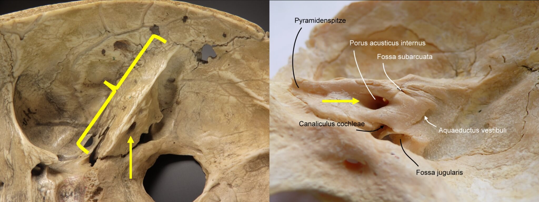
Figure 2
The facialis nerve, after exiting the brain stem, moves to the porus acusticus internus (yellow arrows) and then runs through the petrous bone (yellow bracket) in the bony facialis nerve canal (Canalis nervi facialis) before exiting through a bony opening (Foramen stylomastoideum) at the base of the skull and branching out in the equilateral half of the face.
Source: Modified by author: Welleschik, Own work, Os temporale, pars petrosa, facies posterior, 06/10/2009, commons.wikimedia.org/wiki/File:Ear_internal_anatomy_numbered.svg, accessed on 08/28/2020, CC BY-SA 3.0. creativecommons.org/licenses/by-sa/3.0.
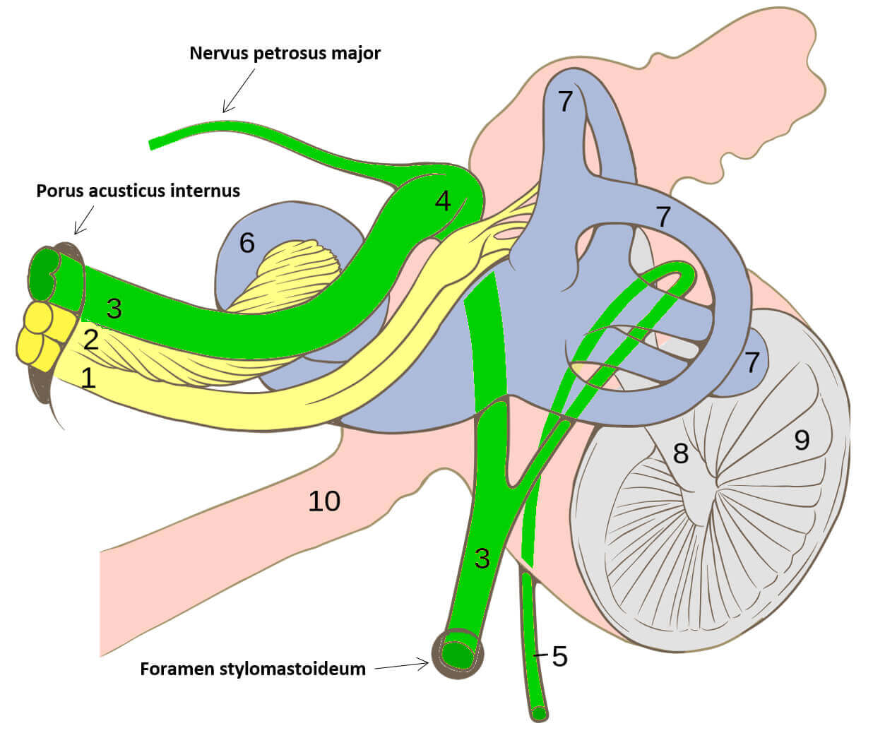
Source: Modified by author: Patrick J. Lynch, numbers by Uwe Gille, Cranial nerves VII and VIII and selected structures of the inner and middle ear. 04/12/2011, commons.wikimedia.org/wiki/File:Ear_internal_anatomy_numbered.svg, accessed on 08/28/2020, CC BY-SA 2.5. creativecommons.org/licenses/by-sa/2.5.
From the brain stem, the facial nerve moves to the opening of the internal auditory canal (Porus acusticus internus, yellow arrows) and then runs through the petrous bone (yellow parenthesis) in the facial nerve canalis, releasing the large petrous nerve (N. petrosus major) to the lacrimal gland, the stapes nerve (N. stapedius) and the tympanic membrane (Chorda tympani) for taste perception and to the salivary glands[4]. The course is shown in the adjacent graph to illustrate the proximity of the numerous structures (common passage of the facial nerve and the nerves for auditory and vestibular perception through the internal acoustic porus) in the temporal bone.
In this model, the course of the facial nerve within the petrous bone (Pars petrosa os-sis temporalis) can be traced in relation to the ossicles (numbers 2 - 4) and to the organ of hearing and balance in the narrower sense (green structure). Furthermore, the two parts of the eighth cranial nerve (N. vestibulochlearis; numbers 8 - 9) are also shown.
Source: Campbell A, Lockwood P, Stewart D, Spielmann P. Anatomy of the Inner Ear. 2015. sketchfab.com/3d-models/anatomy-of-the-inner-ear-f80bda64666c4b8aaac8f63b7b82a0a0. Accessed on 11/15/2020. CC BY-SA 4.0. creativecommons.org/licenses/by-sa/4.0.
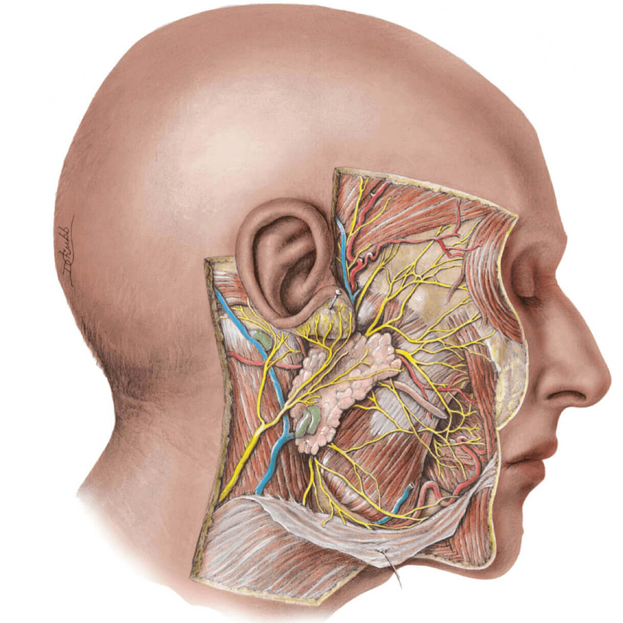
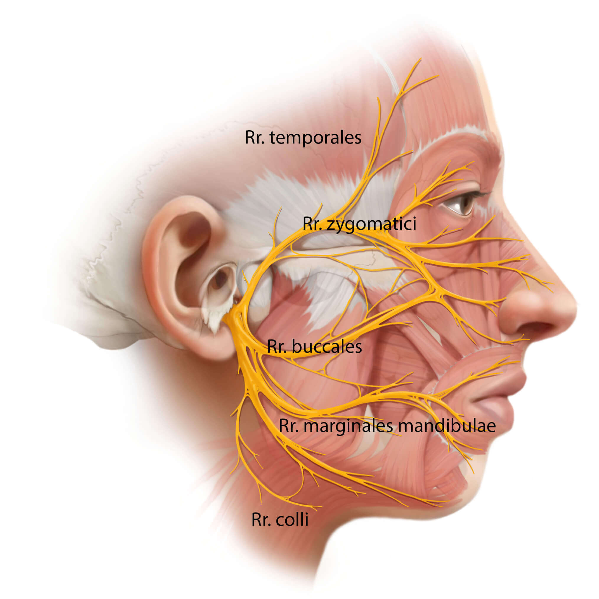
Through the foramen stylomastoideum it emerges from the skull bone and branches in the area of the parotid gland (Glandula parotis), which can be seen in the lower picture as a foamy structure below the ear. The superior branches are called rami (Rr.) (from Latin ramus = branch): Rr. temporales (branches to the temple), Rr. zygomatici (zygomatic branches), Rr. buccales (cheek branches), R. marginalis mandibulae (lower jaw branches) and Rr. colli (branches to the neck). These main branches are further divided into fine lower branches. About 14 branches have already been described on the rostral edge of the parotid gland (towards the mouth)[5].
This 3D-learning model from our friends at ANATOMYNEXT (www.anatomynext.com) shows first of all the N. petrosus major and the Chorda tympani, which both branch off from the N. facialis in the Felsenbein (Pars petrosa ossis temporalis). The N. pe-trosus major serves in particular for the nervous supply of the lacrimal gland (Glan-dula lacrimalis). In contrast, the chorda tympani supplies the mandibular and sub-mandibular glands (Glandulae submandibularis et sublingualis) on the one hand, and on the other hand it enables taste sensations in the front two-thirds of the tongue. The branches of the parotideus plexus are also shown. The parotideus plexus is locat-ed between the superficial and deep part of the parotid gland (Glandula lacrimalis) and innervates especially the mimic muscles. If you click and hold the model with the left mouse button, it can be moved in all directions. The 3D learning model was cre-ated by our friends from ANATOMYNEXT (www.anatomynext.com), who enrich our site with their excellent 2D and 3D teaching models.
The distribution of the branch systems is highly variable (fig. 5) [6]. Furthermore, there are also cross connections among the branch systems, which are of particular interest for synkinesis (explanation under facial paralysis -> forms -> synkinesis) [7, 8].

Source: Alexandra W. Baker, DNA Illustrations, Inc., Lateral view of facial muscles showing six common branching patterns of facial nerve, 222 Flint St.Asheville, NC 28801, n.d., www.dnaillustrations.com/project/facial-nerve-variations, accessed on 08/28/2020.
Source: Modified by author: Alexandra W. Baker, DNA Illustrations, Inc., Lateral view of facial muscles showing six common branching patterns of facial nerve, 222 Flint St.Asheville, NC 28801, n.d., www.dnaillustrations.com/project/facial-nerve-variations, accessed on 08/28/2020.
The 21 pairs of facial muscles allow voluntary and involuntary movements. These also include the platysma and the muscles of the outer ear. The mimetic musculature is divided into four layers, whereby the three superficial layers are innervated from the deep surface and the deepest muscle layer (M. mentalis, M. levator anguli oris, and M. buccinator) are innervated from their superficial surface.
The exact innervation patterns to the facial muscle groups can vary [9]. For example, the muscles that allow mouth movements can also vary in their arrangement [10]. This means that in different people the direction of traction, the amplitude, and the function of the same muscle or the same muscle groups can be quite different.
Sources:
[1] Schmid SM, Hollmann M. Bridging the synaptic cleft: lessons from orphan glutamate receptors. Sci Signal. 2010 Aug 24;3(136):pe28. doi: 10.1126/scisignal.3136pe28. PMID: 20736482.
[2] Corthay J, Dunant Y, Loctin F. Acetylcholine changes underlying transmission of a single nerve impulse in the presence of 4-aminopyridine in Torpedo. J Physiol. 1982 Apr;325:461-79. doi: 10.1113/jphysiol.1982.sp014162. PMID: 6286942; PMCID: PMC1251406.
[3] R. Shane Tubbs, Mohammadali M. Shoja and Marios Loukas. Bergman’s Comprehensive Encyclopedia of Human Anatomic Variation [cited 2018 May 23].
[4] R. Shane Tubbs, Mohammadali M. Shoja and Marios Loukas. Bergman’s Comprehensive Encyclopedia of Human Anatomic Variation [cited 2018 May 23].
[5] Tzafetta K, Terzis JK. Essays on the facial nerve: Part I. Microanatomy. Plast Reconstr Surg 2010; 125(3):879–89.
[6] DAVIS RA, ANSON BJ, BUDINGER JM, KURTH LR. Surgical anatomy of the facial nerve and parotid gland based upon a study of 350 cervicofacial halves. Surg Gynecol Obstet 1956; 102(4):385–412.
[7] Tohma A, Mine K, Tamatsu Y, Shimada K. Communication between the buccal nerve (V) and facial nerve (VII) in the human face. Ann Anat 2004; 186(2):173–8.
[8] Katz AD, Catalano P. The clinical significance of the various anastomotic branches of the facial nerve. Report of 100 patients. Arch Otolaryngol Head Neck Surg 1987; 113(9):959–62.
[9] Kehrer A, Engelmann S, Bauer R, Taeger C, Grechenig S, Kehrer M et al. The nerve supply of zygomaticus major: Variability and distinguishing zygomatic from buccal facial nerve branches. Clin Anat 2018; 31(4):560–5.
[10] Farahvash MR, Abianeh SH, Farahvash B, Farahvash Y, Yagoobi A, Nazparvar B. Anatomic variations of midfacial muscles and nasolabial crease: a survey on 52 hemifacial dissections in fresh Persian cadavers. Aesthet Surg J 2010 [cited 2018 May 23]; 30(1):17–21.