Brow Ptosis (drooping eyebrows)
Often a low level of the brow can be observed due to the slackening of the forehead muscle (Musculus frontalis). This progresses with age and leads to an increasingly clear asymmetry of the face at rest. If the patient contracts the forehead muscle on the unaffected side and lifts the eyebrow there, the inequality of the eyebrow level increases. In order to improve the symmetry whilst at rest, a surgical brow lift can be performed, e.g. by a so-called browpexy or an endoscopic forehead lift (access via small incisions with hidden scars from the crown area).
This 3D learning model from our friends at ANATOMYNEXT (www.anatomynext.com) gives an impression of the relationship between the Galea aponeurotica or Aponeurosis and the M. occipitofrontalis, whose muscle bellies insert in the Galea aponeurotica. In addition, the Mm. auricularis anterior and superior in the ear region insert in the Galea aponeurotica. Together with the scalp (link to skin here), it is also part of the head rind that spans the cranial roof. The 3D learning model was created by our friends from ANATOMYNEXT (www.anatomynext.com), who enrich our site with their excellent 2D and 3D teaching models.
Here it becomes understandable that the sunken brow complex (periorbital complex) results in a limitation of the visual field. The patient reports to see a "black rim" above the right eye in the field of vision towards the "ceiling". The paralyzed frontal muscle as well as other flaccid muscles in the brow complex cause it to sink so that the field of vision is partially obstructed. The remedy is a surgical lifting of the periorbital complex (endoscopic forehead lift).
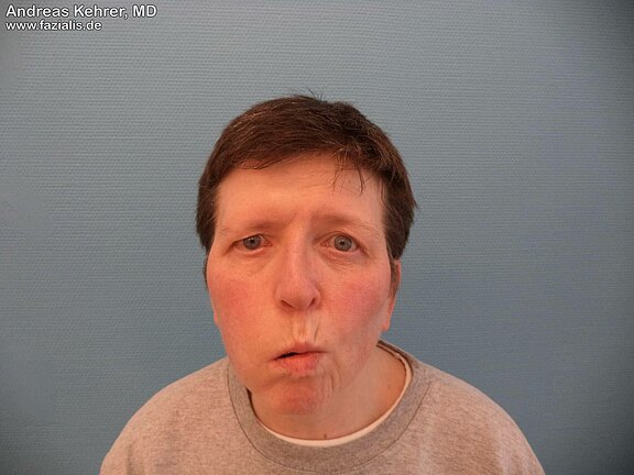
54-year-old female patient with a complete facial paresis on the right side. The flaccid loss of the right forehead muscle is clearly visible. When trying to lift the eyebrows and produce wrinkles on the forehead to an astonished, questioning facial expression, the patient succeeds only on the left side.


