Facial muscles - localisation and function: the mimic muscles (responsible for facial expression) are controlled by the nerve plexus of the facial nerve (see also facial nerve -> mimic musculature). Facial expression is determined by an extremely complex interaction of different individual muscles as well as muscle groups of the mimic musculature and is controlled both arbitrary (conscious) and involuntary (unconscious) movements. The 3D graphic below will afford you a detailed look at the anatomy of the mimic facial muscles from different perspectives. Click on the numbers to see the Latin names of the individual muscles. Zoom in or out with your mouse wheel. Familiarise yourself with the anatomical relationship between individual mimic muscles and other facial structures. Capture the origin and attachment of individual facial muscles to bony protrusions, cartilage structures, and other facial muscles. Try to imagine what happens when there is a shortening of the muscle fibres, i.e. a contraction of the muscle. Which direction of traction is produced? How will mobile neighboring structures react? How will the skin immediately above move? Which wrinkles, which concave and/or convex distortions of the skin are generated by this?
With this 3D learning model from our friends from ANATOMYNEXT (www.anatomynext.com) you can intensively study the facial muscles that are responsible for facial expressions and other important functions. Illustrate the relationship of the individual muscles to each other and to important fixed points in the face, e.g. the eye and mouth complex. Learn the muscle names, their origin (o = origin), insertion (i = insertion), and their individual function (a = action). Zoom/wide-angle works with the mouse wheel. If you click and hold the model with the left mouse button, it can be moved in all directions. The symbol with two arrows at the bottom right shows the model in fullscreen mode. The 3D learning model was created by our friends from ANATOMYNEXT (www.anatomynext.com), who support our site with their excellent 2D and 3D learning models.
| Branch system | Muscle | Function |
|---|---|---|
| Temporal | Eyebrow wrinklers | Pulls eyebrow downward and medially |
| Slim muscle | Pulls medial eyebrow downward | |
| Temporal & Zygomatic | Eye ring muscle | Closes eyelids |
| Zygomatic & Buccal | Large cheekbone muscle | Elevates upper lip |
| Buccal | Cheekbone muscle | Elevates upper lip |
| Upper lip lifter | Elevates upper lip and nasolabial fold | |
| Upper lip and nasal wing lifter | Elevates nasolabial fold and compresses cheek | |
| Risible muscle | Lateral pull on corner of mouth | |
| Cheek muscle | Lateral pull on corner of mouth and compresses cheek | |
| Corner of mouth lifter | Pulls corner of mouth upward and medial | |
| Mouth ring muscle | Closes and compresses lips | |
| Nasal muscle | ||
| Nasal wing lifter | Flares nostrils | |
| Nasal wing closer | Compresses nostrils | |
| Buccal & Marginal mandibular | Mouth angle counterbore | Depresses corner of mouth |
| Marginal mandibular | Lower lip counterbore | Depresses lower lip |
| Chin muscle | Pulls chin upward | |
| Cervical | Cervical muscle | Pulls down jaw, corner of mouth and lower lip |
The opening of the eye and the position of the eyelids is essentially determined by the eye ring muscle (orbicularis oculi muscle). Its origin is located on the, the upper jaw, the eyelid ligament the part of the frontal bone next to the nose. It spans the eye and the eyelids and attaches to the raphe palpebralis lateralis. The eye ring muscle consists of three parts. The so-called pars orbitalis originates from the frontal process of the upper jaw and the nasal part of the forehead bone. The pars palpebralis, on the other hand, originates at the inner eyelid band and is attached to the area of the outer eyelid band. The pars lacrimalis originates from the bone crest of the lacrimal bone (os lacrimale) and surrounds the lacrimal sac.
If the eye ring muscle is relaxed, the eyelid fissure (width of the visible eye, distance between the eyelids) is proportionally wide. If it contracts a little, the eyelid fissure narrows. If its tension is increased consciously or unconsciously, the eyes close slightly. A forced, i.e. a firm eye closure, is achieved by a maximum tension of the eye ring muscle. It can be arbitrary (squinting of the eyes) or involuntary in the context of lightning reflexes (eyelid closure reflex) to protect the eye from damage.
In this model the eye is presented in longitudinal section. At first, the optic nerve and the outer eye muscles can be found in the depth. The external eye muscles can be differentiated into straight and oblique muscles. The superficial parts give an idea of the importance of the facial nerve for closing the eyes, since the orbicularis oculi muscle is innervated by the facial nerve. It should also be emphasized that the apparent hollowness of the eye is filled in vivo, i.e., in the living eye, by the socalled "vitreous body" (Corpus vitreum).
Source: Intervoke. The Human Eye – Cross Section Animation. 2019. sketchfab.com/3d-models/the-human-eye-cross-section-animation-4e7d63d9d3a14ca5aff5f51e01cad153. Accessed on 11/15/2020. CC BY-SA 4.0. creativecommons.org/licenses/by-sa/4.0.
In addition to the eyelid closure function, the eye ring muscle also has other important functions. It is responsible for the even distribution of tear fluid (tear film) from the lacrimal gland, which is located in the outer upper part of the eye socket. The natural blinking reflex is controlled by the facial nerves. Furthermore, the eye ring muscle facilitates the outflow of tear fluid via the tear canal system in the inner corner of the eye.
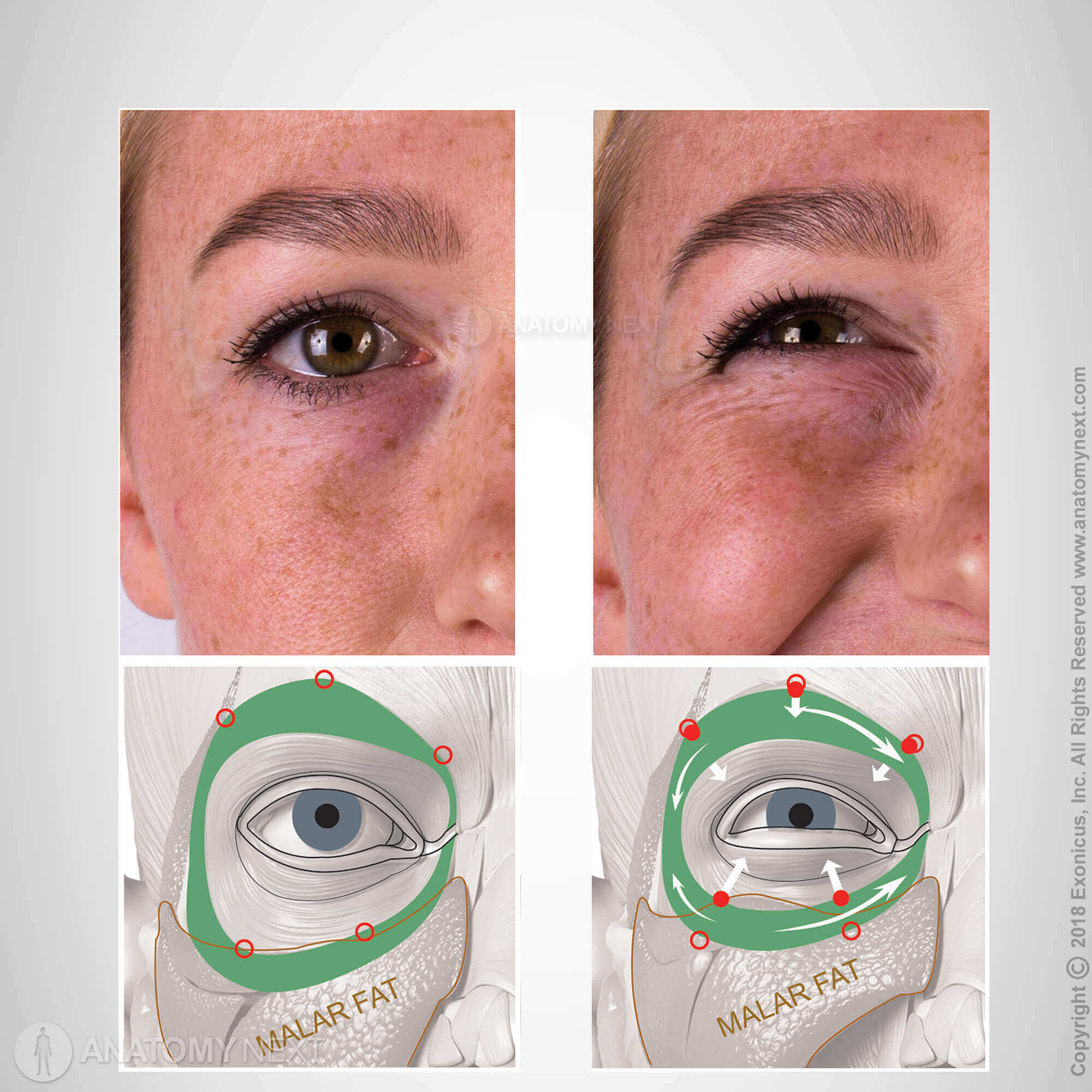
Since the eye ring muscle is joined to the dermis (corium), i.e. it lies directly under the thin layer of skin surrounding the eye, the skin follows every muscle movement. For example, contraction of the muscle at the outer edge of the eye results in typical skin folds, also known as "crow's feet". They become more prominent with increasing age.
Facial Anatomy without Facial Muscles
While the left half of the face shows bony and connective tissue reference points such as the zygomatic bone (Os zygomaticum) or the temporal or temporo-parietal fascia (Fasciae temporalis et temporoparietalis), the right half of the face gives an impres-sion of the complexity of the nervous, articular and venous supply of the face. If you click and hold the left mouse button, the model can be moved in all directions.
Source: Skull Base and Cerebrovascular Laboratory at University of California San Francisco. FTOZ- Model 2. 2018. sketchfab.com/3d-models/ftoz-model-2-b50510ccd1ad43f09e0e6f4d34858b0a. Accessed on 11/15/2020. CC BY-SA 4.0. creativecommons.org/licenses/by-sa/4.0.