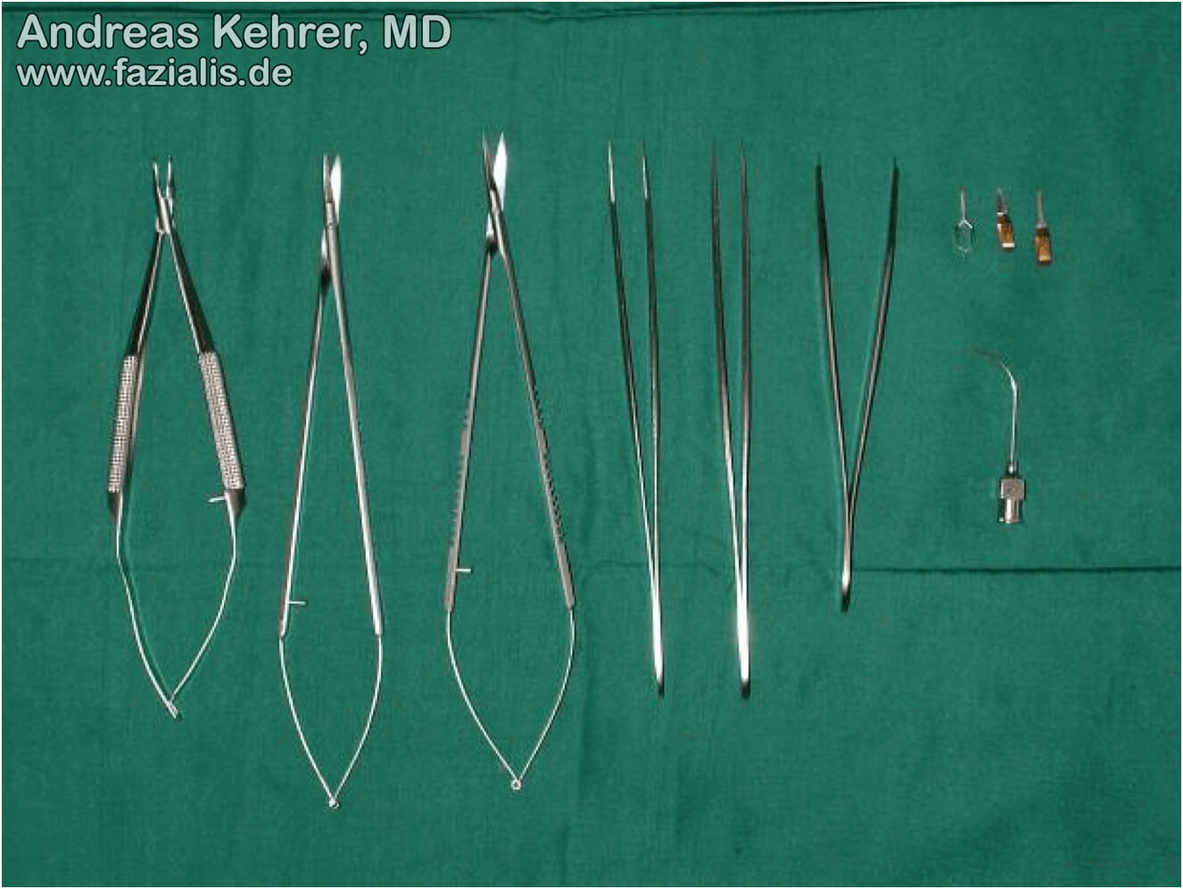
Microsurgery (from Greek "μικρός", "small") is a surgical technique using surgical microscopes or magnifying glasses with high magnification. In the current era, high-resolution special microscopes are used as surgical microscopes which allow a continuous magnification between 10 - 25x. Magnifying glasses suitable for microsurgery have a magnification of 2.5 - 4.5x. For microsurgical operations, a special, particularly fine set of instruments is used. Microsurgical instruments allow the gentle handling of the smallest tissue structures such as the smallest blood vessels (veins and arteries), nerve branches, and nerves. Furthermore, they allow work in places in the human body with very small dimensions.
Sound microsurgical knowledge and skills are indispensable to be able to offer the complete spectrum of reconstructive plastic surgery for facial paralysis. Microsurgical techniques allow for free functional muscle transplants and nerve transfers to be safely performed.
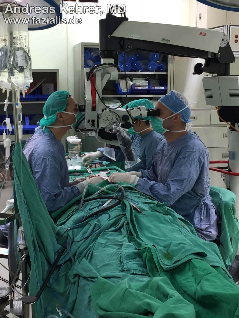
Modern high-performance microscopes with a high magnification factor are used in microsurgery today. The author of this site has trained his microsurgical skills at several internationally renowned centers – here, a picture of a microsurgical head/neck reconstruction at the Chang-Gung-Memorial Hospital (CGMH) in Taipei/Linkou, Taiwan, the world's largest microsurgical center (>1200 free flap procedures/year).
In principle, learning microsurgery does not require any special skills – however, a steady hand, a prudent character, and a lot of patience on the part of the surgeon are of great advantage. Mastering microsurgery requires further prerequisites: in order to be able to offer safe and successful microsurgical operations, they must be performed with constant regularity. Microsurgical sutures of small blood vessels, called "micro-anastomoses", as well as nerve sutures (called "coaptations"), should be learned under all circumstances first in special, certified microsurgical courses and then in animal models, i.e. usually on rats.
In our opinion, a surgeon interested in microsurgery only has the necessary skills to safely and successfully perform the smallest blood vessels on a patient if at least 25 anastomoses have been successfully performed on an animal model. The anastomosis permeability is checked with the so-called "Patency Test", a test procedure during which the blood flow is first spread out with two micro vascular forceps and the blood flow to the blood column is assessed after loosening a pair of forceps.
Sutures with diameter between 8-0 and 12-0 are used. The suture strength and needle size decrease further with increasing number size, i.e. while a 9-0 thread is only slightly finer than a human hair, a 12-0 thread is hardly visible to the naked eye and has about one third the diameter of a human hair.
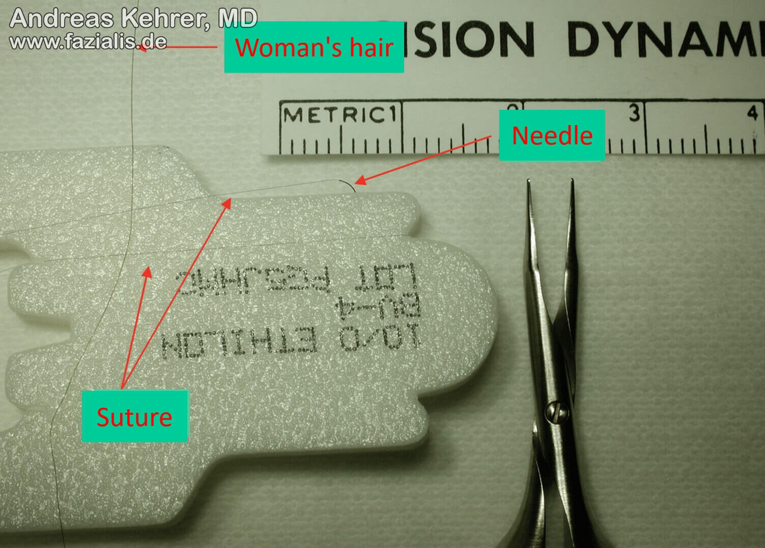
We perform our nerve sutures with a strength of 10-0 in order to introduce as little foreign material as possible for the best nerve healing. The thickness of the thread is extremely fine, so that the suture is hardly visible to the naked eye. It is about one third of a human female hair, as can be seen in the picture above. Precise nerve sutures (coaptations) require advanced microsurgical skills and high-performance microscopes with a high magnification factor.
Transplantation: In the course of a transplant, larger quantities of tissue are usually transplanted from one region of the body to another. The transplanted tissue can only heal in there if it is immediately connected back to the blood supply, i.e. to the blood circulation by microsurgery. Therefore, at least one recipient artery and one donor artery (blood vessel) as well as one recipient vein and one donor vein (blood vessel) must be sutured. For example, if a functional muscle is transplanted, then a recipient nerve is required in the target area, which is connected (coapted) microsurgically to the donor nerve of the muscle transplant in order to control the muscle. Free tissue transplants are used in plastic surgery when their native function can consciously be dispensed without any relevant disadvantage. In the case of free functional muscle transplantation, such as gracilis muscle transfer from the thigh, surrounding muscles automatically take over the function. A free functional toe transplantation as thumb replacement also functions according to this principle: if, for example, the 2nd toe is "sacrificed" for this, the remaining toes take over important functions for statics and gait pattern.
Replantation: If limbs or parts of the body are separated, e.g. by accidents, they can be "replanted" with the help of microsurgery, provided that no extensive bruising is present. Microreplantation is the term used when amputations involve smaller parts of the body, such as fingers in the context of circular saw injuries. Macroreplantations are larger parts of the body and limbs that are reattached with the surgical microscope, such as cuts in the lower limb at lower or thigh level, which are provided with a microsurgical restoration of blood and nerve supply to maintain function.
Micro repair: Micro repair is the term used to describe the microsurgical restoration of severed nerves and vessels (e.g. as a result of an accident or as part of a tumor resection).
Professionalism in microsurgery is not defined solely by the ability to perform a successful microsurgical anastomosis in the laboratory or on the patient - in the case of a flap transplantation, it includes the reliable indication, selection of the tissue transplant, selection of the required tissue components, selection and isolation of suitable connecting vessels and, if necessary, nerves, rapid and safe transplantation of the tissue, assessment of the perfusion of the flap tissue (1) as well as the planning of any necessary secondary operations. Microsurgery is used, among other things, in interventions on small blood vessels, the central nervous system, the lymphatic system, and in peripheral nerve surgery.


In the context of microsurgery courses and training models, there is no excuse for the microsurgeon for suboptimal performance. Even if the vessels demonstrated the desired permeability of the anastomosis by means of a "patency test" and external supervision, they are opened completely after completion of the exercise and the distance between the stitches, the stitch depth, the strength of the sutures and knots, the quality of the vascular wall adaptation, and other criteria are checked in their consistency along the microseam row under the microscope. Vascular tears should be just as invisible as possible micro blood clots along the anastomosis. This "bypass" of the author on the rat model, with the main neck artery (carotid artery) attached to the main abdominal artery (aorta) with the finest 11-0 suture material, fulfilled all required criteria (from the "Micro-Lab" training laboratory of plastic surgery at the Erasmus Medical Center/Rotterdam).