Source: Modified by author: Patrick J. Lynch, Dr. C. Carl Jaffe, Cranial nerve IX 9 Bell's palsy, 12/23/2006, commons.wikimedia.org/wiki/File:Cranial_nerve_VII_bells_palsy.svg, accessed on 08/28/20, CC BY-SA 2.5. creativecommons.org/licenses/by-sa/2.5.
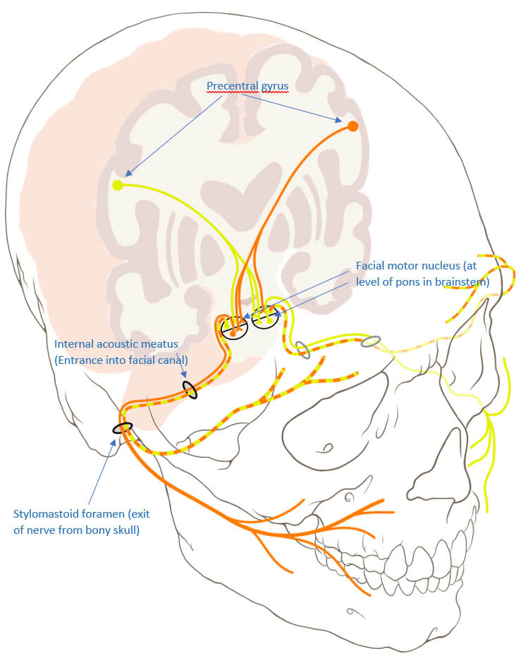
Physiology of facial expression at the cerebral level: each movement of the mimetic musculature is planned in the cerebral cortex (in the region of the Precentral Gyrus) and sent via a path (Fibrae corticonucleares) to the nuclei of the facial nerve located in the brain stem. These consist of a divided cell group. The upper cell group supplies the eyelid and forehead muscles and is controlled by both brain hemispheres (bilaterally). The lower nuclear cell group, which is responsible for the innervation of the remaining mimetic muscles, is only controlled by crossed fibers of the opposite (contralateral) side (unilaterally). This allows a differentiation between a central and a peripheral paralysis of the facial nerve, as in central paralysis frowning may still be possible on the affected side.
This model allows you to visualize the macroscopic structure of the brain. The two halves of the brain (hemispheres) are separated by a central furrow (Fissura longitudinalis). Furthermore, the brain convolutions (Gyri cerebri) and the notches between them (Sulci cerebri) are shown. Furthermore, one can differentiate between anterior (Prosencephalon), middle (Mesencephalon) and the so-called "rhombic brain" (Rhombencephalon). In the brain stem, more precisely in the so-called "bridge" (Pons), the control nuclei of the N. facialis are located. The spinal cord (Medulla spinalis) is attached to them. On average, the human brain weighs 1.300 to 1.600 grams.
Source: Skull Base and Cerebrovascular Laboratory at University of California San Francisco. CPs - Model 1: Sulci & Gyri of Left Hemisphere. 2020. sketchfab.com/3d-models/cps-model-1-sulci-gyri-of-left-hemisphere-216d50de44084049800ac0d548fb51b5. Accessed on 11/15/2020. CC BY-SA 4.0. creativecommons.org/licenses/by-sa/4.0.
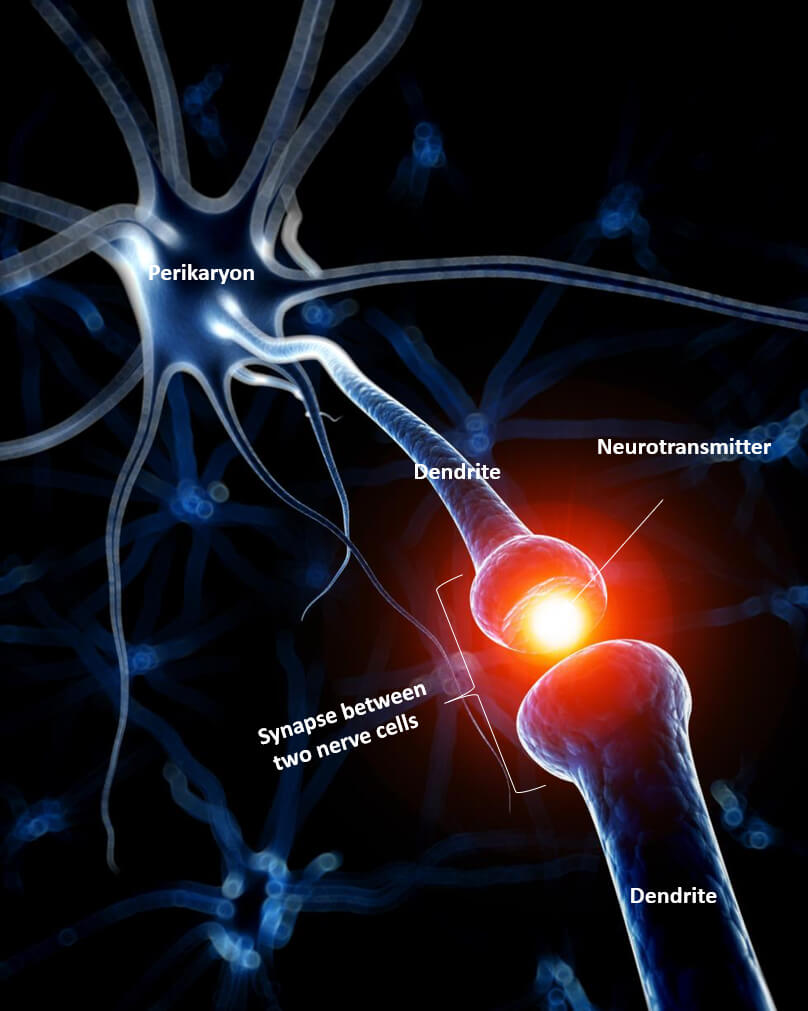
The information, or electrical impulse, is transmitted from the brain via the nerves, which consist of many neurons, to the muscle. The upper part of the picture shows the cell body containing the nucleus. The cell body is surrounded by so-called dendrites, small appendages which serve the excitation reception of the nerve cell. The long part in the middle is called the axon. The axon serves the transmission of excitation and makes up a large part of the macroscopically visible nerve in the sum of many nerve cells. Peripheral facial paresis leads to partial damage or even complete interruption of the continuity of the axon (and neighboring axons). The extensions of the axon, in the lower part of the picture, are motor end plates. They transmit the stimulus to mimetic muscle cells.

This graphic illustrates the components of a nerve cell (neuron) as shown in Fig. 2. Note in particular the numerical ratio of dendrites to axon. While a neuron can receive stimuli via a large number of dendrites, only one axon with a variety of endings, so-called "boutons", is available for transmitting stimuli.
Source: Versal. Neuron. 2016. sketchfab.com/3d-models/neuron-d40557a1e4154267b78117433bc51296. Accessed on 11/15/2020. CC BY-SA 4.0. creativecommons.org/licenses/by-sa/4.0.
Here the process of stimulus absorption is illustrated via the dendrites to the nerve cell body (soma). The electrical excitation propagation of the dendrites is afferent, i.e., directed towards the cell body. Dendrites themselves can also carry so-called "dendritic spines" (Spinulae dendriticae). These protrusions are between 0.2 and 2 µm long. In particular mushroom-shaped dendritic spines can also serve as calcium stores and influence synaptic plasticity.
Source: Neural Impulse Media. Neuron Action Potential. 2015. sketchfab.com/3d-models/neuron-action-potential-c7ca6da31f5848748f7ba7ccb2508266. Accessed on 11/15/2020. CC BY-SA 4.0. creativecommons.org/licenses/by-sa/4.0.
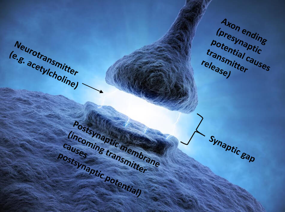
Computer schematic drawing of a motor end plate, also known as a neuromuscular endplate. It transmits the excitation from a nerve fiber to the muscle fiber and represents a chemical synapse. This means that the transmission is transmitted via messenger substances such as acetylcholine (a neurotransmitter). In contrast to muscle fibres in the rest of the body, several motor end plates are located on a mimetic muscle fibre[1]. Botulinum toxin (Botox R) is a neurotoxin which is also used as an effective medication. It inhibits the release of the neurotransmitter acetylcholine. If desired, the muscle function can be reduced or completely blocked. Botulinum toxin is used, for example, in spastic forms of facial paresis or synkinesis.
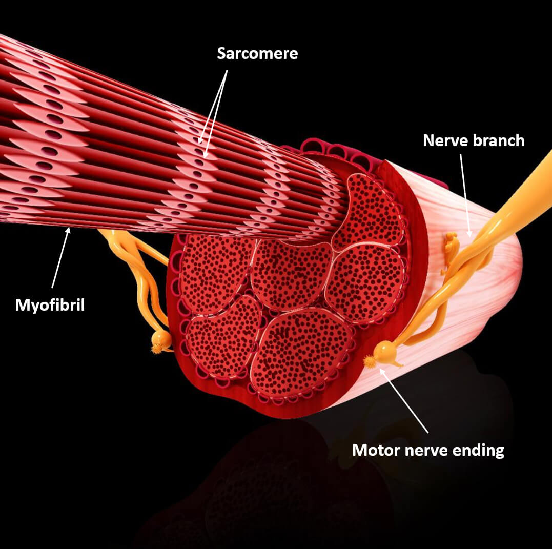
Muscle fibers contain one to several hundred muscle fibrils (myofibrils), which are all aligned in parallel. The muscle fibrils contain sarcomeres, which form the contractile units of the muscle cell. They have a certain resting voltage (resting tone). If they are now excited more strongly, the sarcomeres are shortened, whereby the entire muscle is shortened and mimic movement occurs. The largely unconsciously-controlled coordination of many individual movements of the different facial muscles finally leads to facial expression.
The muscle contraction can be differentiated according to an isotonic or isometric pattern. In the former, the muscular tension is maintained while the muscle length changes; vice versa in the isometric form. A change in muscle length can be concentric in the form of a shortening or, conversely, eccentric. Strength training, for example, can achieve muscular hypertrophy, i. e. an increase in muscle size by increasing the muscle cell volume. Such hypertrophy can be caused in the facial muscles by excessive teeth grinding (bruxism).
Source: "kmith". Muscle Contraction. 2019. sketchfab.com/3d-models/muscle-contraction-f2f6660073e3429aad069f49fbe2eb5b. Accessed on 11/15/2020. CC BY-SA 4.0. creativecommons.org/licenses/by-sa/4.0.
In reflexes, the facial nerve is activated by stimulation of other nerves. This is the case with the blinking reflex, which is triggered by the trigeminal nerve (the fifth cranial nerve). The blinking reflex is triggered by an irritation of the first trigeminal branch (N.supraorbitalis) by touching the eyelashes. This information is passed on to the facial nerve in the brain stem. The zygomatic facial nerve branches then trigger a short bilateral contraction of the eye ring muscle (orbicularis oculi muscle), while other muscle groups, such as the mouth ring muscle (orbicularis muscle), are not activated. Thus, the facial nerve mediates an important protective reflex.