The facial nerve, with its branch systems, is responsible for controlling the facial muscles (mimetic muscles). The external shape of the face is mainly characterized by bony structures (bony eye socket, cheekbone, lower jawbone), but it acquires its individual character and expression through the mimetic musculature. Furthermore, depressions such as the chin-lip furrow (Sulcus mentolabialis) and the nasal cheek furrow (Sulcus nasolabialis) contour the face. The 21 muscles in each half of the face react in fractions of a second and can reflect emotions in an extremely varied way. This is possible due to the special conception of these muscles. They lie without fascia (connective tissue of skeletal muscles) in the subcutaneous fatty tissue layer, in which they are anchored via elastic tendons. Another special feature is the increased number of motor end plates on the muscle fibres.
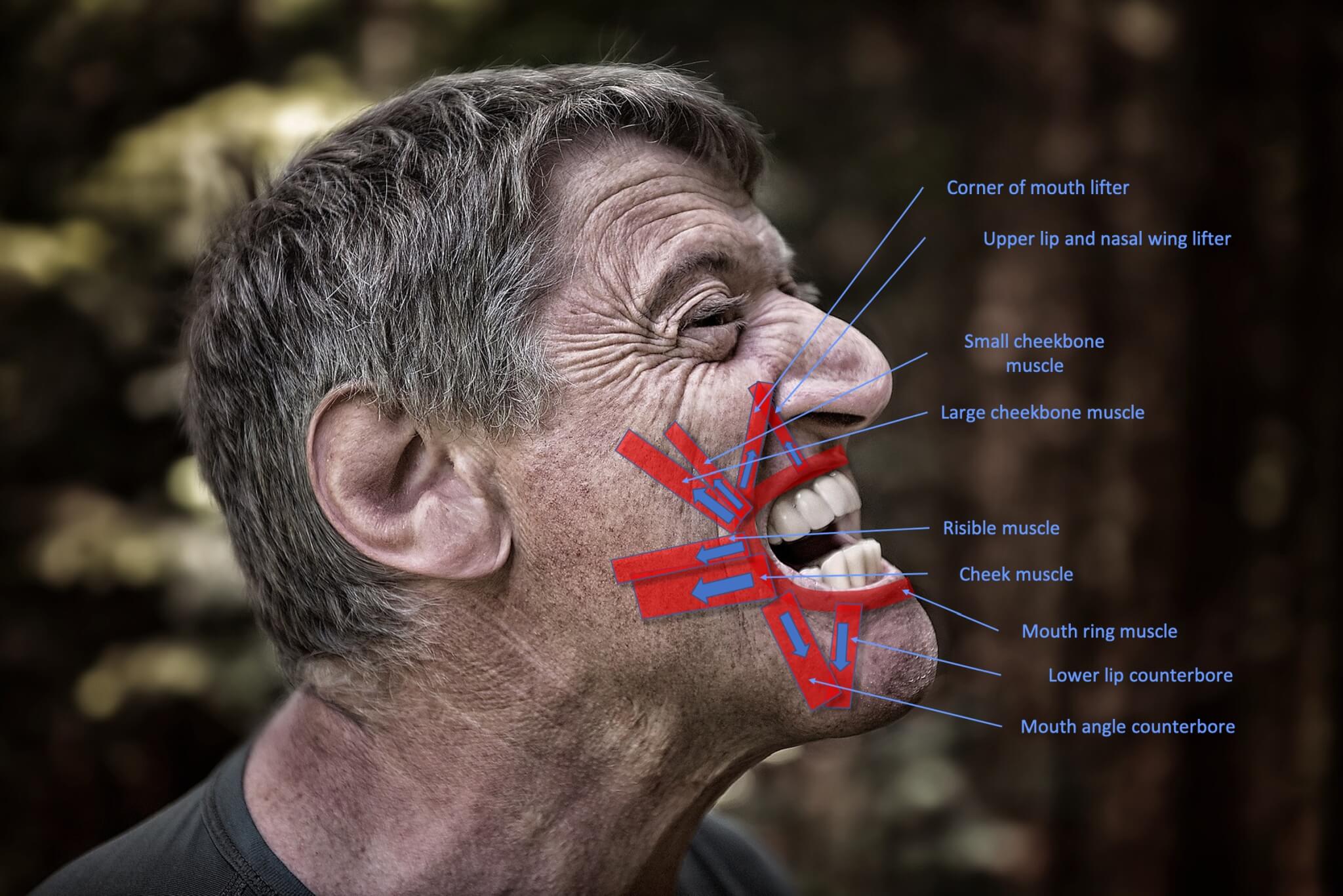
In comparison to skeletal muscles, where there is only one endplate per fiber, which is usually located centrally, mimetic muscles usually have several motor end plates distributed over the entire fiber course [1]. The facial muscles can thus be controlled more precisely and dynamically. Two essential mimetic muscles are the eye and mouth ring muscles. The eye ring muscle (orbicularis oculi muscle) is located directly under the skin around the eye. It has an important influence on the function and facial expression of the eyelids. The mouth ring muscle (orbicularis oris muscle), on the other hand, sometimes controls the width and configuration of the mouth opening. Ifit is intentionally contracted completely, the lips become pointed. Both muscles are schematically shown in the photo above right. The muscles shown in the lower picture, the mouth and cheek area, are attached to the corner of the mouth and the ring muscle at different points and also have different tensile directions. This interaction of the vectors
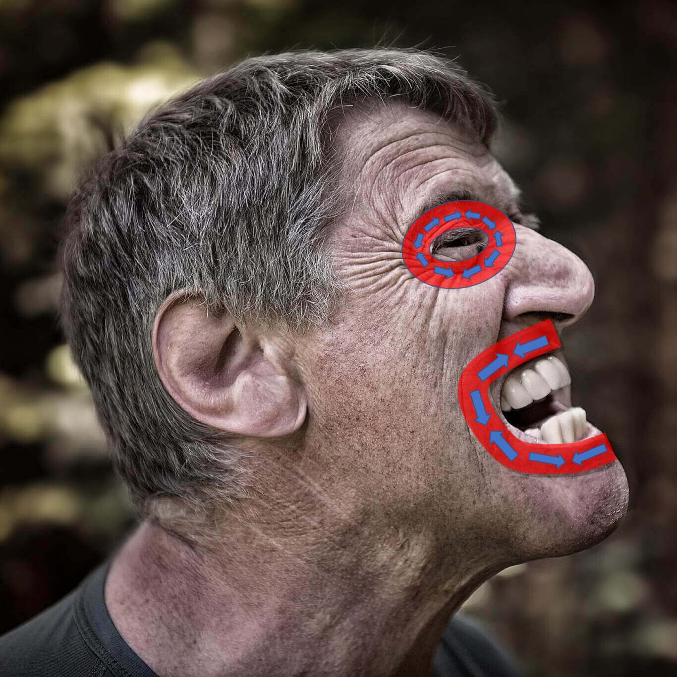
Makes the mouth opening and the smile multi-faceted. Furthermore, the insertion and main vector direction of the muscles vary from person to person, especially in the region of the mouth and cheek the variations are large [2]. The interaction of the muscles can be studied with the help of the animations below.
| Branch system | Muscle | Function |
|---|---|---|
| Temporal | Eyebrow wrinklers | Pulls eyebrow downward and medially |
| Slim muscle | Pulls medial eyebrow downward | |
| Temporal & Zygomatic | Eye ring muscle | Closes eyelids |
| Zygomatic & Buccal | Large cheekbone muscle | Elevates upper lip |
| Buccal | Cheekbone muscle | Elevates upper lip |
| Upper lip lifter | Elevates upper lip and nasolabial fold | |
| Upper lip and nasal wing lifter | Elevates nasolabial fold and compresses cheek | |
| Risible muscle | Lateral pull on corner of mouth | |
| Cheek muscle | Lateral pull on corner of mouth and compresses cheek | |
| Corner of mouth lifter | Pulls corner of mouth upward and medial | |
| Mouth ring muscle | Closes and compresses lips | |
| Nasal muscle | compresses dorsum of nose, dilates nostrils | |
| Nasal wing lifter | Flares nostrils | |
| Nasal wing closer | Compresses nostrils | |
| Buccal & Marginal mandibular | Mouth angle counterbore | Depresses corner of mouth |
| Marginal mandibular | Lower lip counterbore | Depresses lower lip |
| Chin muscle | pushes central lip upward, pulls chin upward | |
| Cervical | Cervical muscle | Pulls down jaw, corner of mouth and lower lip |
With this 3D learning model you can intensively study the facial muscles that are responsible for facial expressions and other important functions. Illustrate the relationship of the individual muscles to each other and to important fixed points in the face, e.g. the eye and mouth complex. The 3D learning model was created by our friends from ANATOMYNEXT (www.anatomynext.com), who support our site with their excellent 2D and 3D learning models. Learn the muscle names, their origin (o = origin), insertion (i = insertion), and their individual function (a = action). Zoom/wide-angle works with the mouse wheel. If you click and hold the model with the left mouse button, it can be moved in all directions. The symbol with two arrows at the bottom right shows the model in fullscreen mode.
| branch system | muscle | function |
|---|---|---|
| Temporal | Corrugator supercilii | Pulls eyebrow downward and medially |
| Procerus | Pulls medial eyebrow downward | |
| Temporal & Zygomatic | Orbicularis oculi | Closes eyelids |
| Zygomatic & Buccal | Zygomaticus major | Elevates upper lip |
| Buccal | Zygomaticus minor | Elevates upper lip |
| Levator labii superioris | Elevates upper lip and nasolabial fold | |
| Levator labii superioris alaeque nasi | Elevates nasolabial fold and compresses cheek | |
| Risorius | Lateral pull on corner of mouth | |
| Buccinator | Lateral pull on corner of mouth and compresses cheek | |
| Levator anguli oris | Pulls corner of mouth upward and medial | |
| Orbicularis oris | Closes and compresses lips | |
| Nasalis | compresses dorsum of nose, dilates nostrils | |
| Dilator naris | Inflates nostrils | |
| Compressor naris | Compresses nostrils | |
| Buccal & Marginal mandibular | Depressor anguli oris | Depresses corner of mouth |
| Marginal mandibular | Depressor labii inferioris | Depresses corner of mouth |
| Mentalis | pushes central lip upward, pulls chin upward | |
| Cervical | Platysma | Pulls down jaw, corner of mouth & lower lip |
Facial Anatomy without Facial Muscles
While the left half of the face shows bony and connective tissue reference points such as the zygomatic bone (Os zygomaticum) or the temporal or temporo-parietal fascia (Fasciae temporalis et temporoparietalis), the right half of the face gives an impression of the complexity of the nervous, articular and venous supply of the face. If you click and hold the left mouse button, the model can be moved in all directions.
Source: Skull Base and Cerebrovascular Laboratory at University of California San Francisco. FTOZ- Model 2. 2018. sketchfab.com/3d-models/ftoz-model-2-b50510ccd1ad43f09e0e6f4d34858b0a. Accessed on 11/15/2020. CC BY-SA 4.0. creativecommons.org/licenses/by-sa/4.0.
Sources:
[1] Happak W, Burggasser G, Liu J, Gruber H, Freilinger G. Anatomy and Histology of the Mimic Muscles and the Supplying Facial Nerve. In: Stennert ER, Kreutzberg GW, Michel O, Jungehülsing M, editors. The Facial Nerve. Berlin, Heidelberg: Springer Berlin Heidelberg; 1994. p. 85–6.
[2] Farahvash MR, Abianeh SH, Farahvash B, Farahvash Y, Yagoobi A, Nazparvar B. Anatomic variations of midfacial muscles and nasolabial crease: a survey on 52 hemifacial dissections in fresh Persian cadavers. Aesthet Surg J 2010 [cited 2018 May 23]; 30(1):17–21.